MRI Technology: The Remarkable Scientific Journey of Nuclear Magnetic Resonance Unlocking the Secrets of the Human Body
Discover how magnetic fields and radio waves are used to obtain highly accurate images of internal organs without X-rays.
Magnetic Resonance Imaging (MRI) is a revolutionary invention in modern medical science that has given doctors the remarkable power to look inside the body without cutting it open. Used in radiology, this technique utilizes powerful magnetic fields, magnetic field gradients, and radio waves to create detailed images of the body's anatomy and physiological processes. The greatest feature of MRI is that it does not use X-rays or ionizing radiation, which distinguishes it from computed tomography (CT) and positron emission tomography (PET) scans. This technique is fundamentally based on the principle of nuclear magnetic resonance (NMR), which is also used in other branches of science to determine the structure of substances.
The Fascinating Journey of History and Nomenclature
The credit for discovering nuclear magnetic resonance goes to Isidor Rabi, who first measured this phenomenon in a molecular beam in 1938. He was awarded the Nobel Prize in Physics in 1944 for this significant work. Subsequently, in 1946, Felix Bloch and Edward Mills Purcell extended this technique to liquids and solids, for which they received the Nobel Prize in 1952. In the early 1970s, R. Damadian, C.F. Hazlewood, and D.C. Chang demonstrated that NMR relaxation times could be used to detect cancer. Soon after, P.C. Lauterbur proposed using magnetic field gradients to form images in a 2D plane.
Initially, this technique was known as NMRI (Nuclear Magnetic Resonance Imaging). However, to eliminate the negative fear associated with the word 'nuclear', this word was later dropped from it, and only the name MRI became popular. Today, approximately over 50,000 MRI scanners are operational worldwide, making them indispensable in hospitals for disease diagnosis and treatment planning.
- Magnetic AlignmentHydrogen protons in the body are aligned in one direction by a powerful static magnetic field (B0).
- Radio Pulse ExcitationThe alignment of protons is disrupted by emitting a radio frequency pulse of a specific frequency.
- Signal DetectionWhen the pulse stops, protons return to their original state and emit radio signals, which are captured by detection coils.
- Image GenerationA computer analyzes these signals to create detailed 2-D or 3-D images of the internal parts of the body.
How Does MRI Work? The Secret Behind the Science
A large part of the human body is made up of water and fat, in which hydrogen atoms are abundant. An MRI scan primarily maps the location and behavior of these hydrogen atoms present in the body. The hydrogen nucleus contains only one proton, which naturally spins on its axis and acts like a tiny magnet. When a patient is placed inside the powerful magnetic field of an MRI machine, these protons align in the direction of the magnetic field. This process is called polarization.
Once the protons are aligned, the machine emits a radio frequency (RF) pulse of a specific frequency. This energy disrupts the alignment of the protons and deflects them from their original state. When the RF pulse is turned off, the protons return to their original state. During this return process, they emit radio signals, which are captured by the antennas (RF coils) inside the machine. These signals are processed by a computer and converted into highly detailed images.
| Type | Capacity (in Tesla) | Main Usage |
|---|---|---|
| Portable/Open MRI | 0.2 to 1.0 T | Claustrophobic patients and bedside scanning |
| Standard Clinical | 1.5 T | General hospital diagnosis and routine scan |
| Advanced Clinical | 3.0 T | High-quality images of brain and soft tissues |
| Research | 7.0 to 11.7 T | Neuroscience and extremely complex medical research |
Internal Structure of the Machine and the Echoing Sound
The main components of an MRI scanner include the main magnet, shim coils, gradient system, and RF system. The main magnet is extremely powerful, with its strength measured in units of Tesla (T). Most clinical systems operate at 1.5 T or 3 T. These magnets are typically superconducting, and liquid helium is used to keep them extremely cold. The loud noise heard during a scan is caused by the gradient coils. These coils rapidly change the magnetic field, causing a process called magnetostriction, which creates noise due to slight vibrations in the machine's wiring.

T1 and T2 Relaxation: Understanding the Difference
Image clarity and contrast in an MRI depend on two main processes: T1 and T2 relaxation. T1 relaxation, also known as spin-lattice, is the time it takes for protons to realign with the direction of the magnetic field. This type of image is excellent for identifying fatty tissues and obtaining structural information about the body. On the other hand, T2 relaxation, or spin-spin relaxation, is the time in which the coherence among protons decays. T2-weighted images are very useful for detecting swelling (edema), inflammation, and the presence of fluid in the body. Because the T1 and T2 values of water and fat are different, doctors can clearly distinguish between various tissues.
Tesla (T)
The Role of MRI in Medical Diagnosis
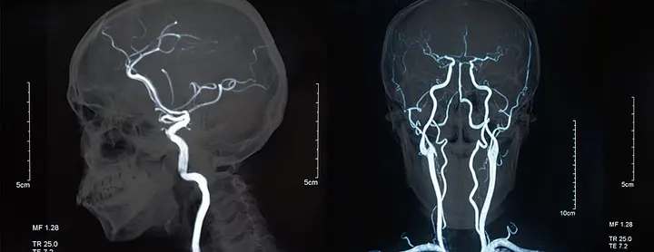
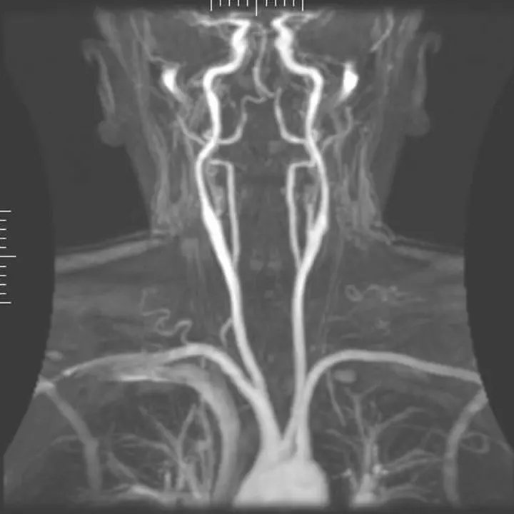
MRI is considered the premier tool for neuroimaging, which is the examination of the brain. It is extremely accurate in diagnosing conditions such as brain tumors, multiple sclerosis, stroke, and Alzheimer's disease. Compared to a CT scan, an MRI provides much better images of the posterior fossa of the brain. In addition, it is widely used for heart diseases, muscle and joint injuries, and cancer staging. Magnetic Resonance Angiography (MRA) allows for the observation of blood flowing through blood vessels, which is helpful in investigating heart attacks or blockages.
Safety and Precautions
Although MRI is safe, certain precautions are essential due to its powerful magnetic field. An MRI can be dangerous for patients with pacemakers, metal implants, or other non-removable metal objects in their body. These metal objects can be pulled toward the magnet or become heated. Furthermore, due to the closed tube-like structure, some patients experience claustrophobia (fear of enclosed spaces), to solve which 'open' MRI machines are now also available. In 2020, the FDA also approved a portable MRI scanner, which uses a lower-capacity magnet.

Future Direction and Artificial Intelligence
MRI technology is now turning toward Artificial Intelligence (AI). Collaborations such as the 'fastMRI' project between Meta AI Research (FAIR) and NYU Langone Health have demonstrated that with the help of AI, the scanning time can be reduced by 4 times. This will require patients to spend less time in the machine and allow hospitals to scan more patients. In the field of research, ultra-high-field magnets ranging from 7 T to 11.7 T are now being used, which will show brain function with a clarity never seen before. In the future, this technology is expected to become more affordable and accessible, making diagnosis much easier for the general public.