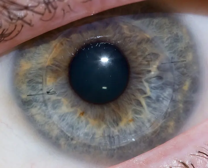
A Medical Revolution in Ocular Restoration: The Science of Corneal Transplants and Vision Recovery
An in-depth look at how modern science restores vision through donor tissue or artificial alternatives when the natural lens becomes opaque.
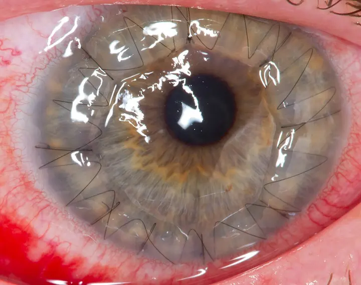
The eye is the most complex and sensitive organ of the human body, connecting us to the outside world. From a scientific perspective, the eye is a sensory system that detects light and converts it into electro-chemical impulses in neurons. According to Wikipedia reports, in higher organisms, the eye is a complex optical system that collects light from the surrounding environment and focuses it through a lens to form an image on the retina. The most important part in this process is played by the 'cornea', which is the outermost transparent part of the eye.
The cornea acts like a protective window covering the pupil, iris, and lens of the eye. When this window becomes cloudy or its shape is distorted due to disease, injury, or genetic factors, an individual's visual ability is severely affected. In such situations, 'corneal transplantation' or 'keratoplasty' emerges as a ray of hope. This is a surgical procedure in which the damaged cornea is replaced by healthy corneal tissue, or a graft, obtained from a deceased donor.
The Need and Purposes of Corneal Transplant
Corneal transplants are primarily performed for four reasons. The first and most common reason is optical, where opaque or distorted tissue is replaced to improve vision. This includes conditions like keratoconus, in which the shape of the cornea becomes cone-like. The second reason is tectonic or reconstructive, which aims to maintain the anatomy of the eye, especially if the cornea has become thin or has developed a perforation.
The third reason is therapeutic, in which inflamed or infected tissue that does not respond to antibiotics is removed. Finally, this surgery can also be performed for cosmetic reasons, to remove white scars on the cornea and improve the appearance of the eye. According to Source 1, as of 2021, the success rate of this procedure has been recorded at a minimum of 41%. This surgery is performed by ophthalmologists, who are eye specialist doctors.
- AnesthesiaThe eye is numbed by administering local or general anesthesia to the patient.
- TrephinationA circular piece of the damaged cornea is cut and removed using an instrument called a trephine.
- Graft placementA piece of the same size is cut from the donor's healthy cornea and placed in the patient's eye.
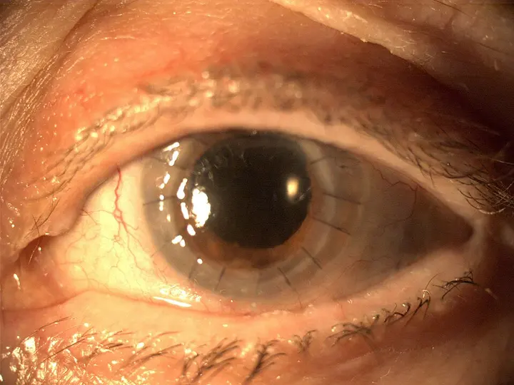
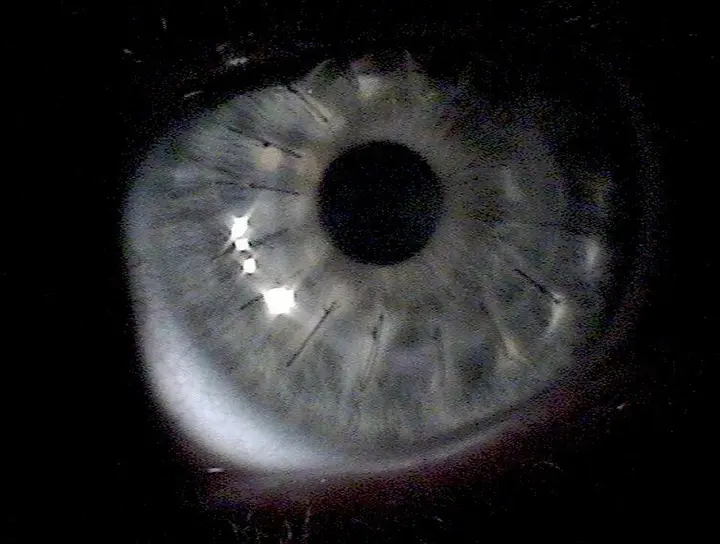
- SuturingThe new cornea is securely stitched into place using extremely fine sutures.
Types of Surgery: Penetrating versus Lamellar
There are two main types of keratoplasty: penetrating and lamellar. When the entire cornea is replaced, it is called penetrating keratoplasty. In this method, a circular piece of the patient's cornea is removed using a circular cutting instrument called a trephine, and it is replaced with an equal portion from a donor, which is then secured with sutures, or surgical stitches.
On the other hand, in lamellar keratoplasty, only certain layers of the cornea are replaced. Since the total thickness of the cornea is only 500 μm (micrometers), this procedure is extremely challenging. The main advantage of this method is that the structural integrity of the eye is maintained. This includes techniques such as 'Deep Anterior Lamellar Keratoplasty' (DALK), in which the upper layers of the cornea are replaced while the inner endothelium layer is kept intact.
સ્નેલન ચાર્ટ રેશિયો
Endothelial Keratoplasty: A Modern Revolution
In the last few years, endothelial keratoplasty has revolutionized the treatment of corneal diseases. In this procedure, only the innermost layer of the cornea, called the endothelium, is replaced. Methods such as DSEK and DMEK are popular in this regard. In DSEK, the donor tissue is 100-150 μm thick, whereas in DMEK, only the endothelium is transplanted.
The biggest advantage of this method is that it requires no stitches or very few stitches. Patients can regain their vision in just a few days or weeks, whereas a full transplant can take up to a year. During surgery, an air bubble is placed inside the eye to hold the new tissue in place, which helps the tissue adhere naturally. However, according to an Australian study, the loss of endothelial cells in DSEK can be higher than in a full transplant.
CAIRS: New Hope for Keratoconus
In 2018, a new technique called 'Corneal Allogenic Intrastromal Ring Segments' (CAIRS) was introduced by Dr. Soosan Jacob. This method is specifically for patients with keratoconus. In this, instead of plastic, small pieces made from donor corneal tissue are inserted between the layers of the cornea. Being a biological material, the possibility of infection or rejection by the body remains low.
The CAIRS technique is extremely flexible because the graft can be customized according to the shape of each patient's eye. Experts based in Brisbane have also created a free website, cairsplan.com, for planning this surgery. This method is now officially recognized in countries like Australia, the UK, and Japan. It strengthens the weakened cornea and significantly improves visual clarity.
Surgical Procedure and Precautions
On the day of the surgery, the patient is brought to the hospital or an outpatient center. The procedure is generally performed under local anesthesia and sedation, so that the patient is awake but does not feel pain. In the case of children, general anesthesia is administered. A lid speculum is used during the surgery to keep the eye open.
After surgery, the patient is given antibiotic eye drops and the eye is covered with a bandage. The cornea does not have blood vessels, so it heals more slowly than other parts of the body. During this period, there is a risk of infection, so it is extremely important to administer the drops according to the doctor's advice. In most cases, the patient is called for a check-up the very next day after surgery.
- PreparationThe area around the eye is prepared by administering local or general anesthesia to the patient.
- Removal of damaged tissueThe damaged part of the patient's cornea is cut and separated in a circular manner using a trephine instrument.
- Graft transplantationThe healthy corneal tissue obtained from a donor is placed in the same spot and stitched using surgical sutures.
Risks and Graft Rejection
Like any surgery, there are risks involved in a corneal transplant. The biggest risk is graft rejection, which is seen in about 10% of cases. This means that the patient's body does not accept the new tissue and attacks it. This reaction can occur even years after the surgery. To prevent this, immunosuppressants like cyclosporine A are used.
Another risk is the transmission of infectious diseases, although it is extremely rare. All donor tissues are strictly tested for viruses like HIV and hepatitis. Interestingly, since 1994, there has been a ban in the United States on the donation of corneas by men who have had physical relations with another man in the last 5 years, even if their HIV report is negative. This policy is still in effect there today.
Artificial Cornea: Boston Keratoprosthesis
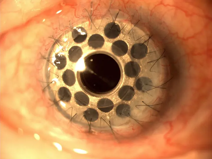
When natural corneal transplants fail repeatedly or are not possible, an artificial cornea is used. Among these, the 'Boston keratoprosthesis' is the most well-known. By 2008, more than 900 such surgeries had been performed worldwide. This is a synthetic device that replaces the damaged cornea and allows light to enter the eye.
In a country like India, where the rate of corneal blindness is high, this technology and awareness about organ donation are very important. Donors can be of any age, such as a 10-year-old child named Janice Babson who donated her eyes after death. Through the confluence of modern science and human generosity, millions of people today have become able to see the world again.