MRI Technology: Mapping the Internal Structure of the Human Body Using Magnetic Fields and Radio Waves
A unique convergence of quantum physics and medical science, MRI provides high-resolution imaging of soft tissues without the use of harmful radiation. An in-depth exploration of this technology.

Magnetic Resonance Imaging (MRI) is a sophisticated medical imaging technique used in the field of radiology that produces highly clear images of the body's internal organs and physiological processes. This system uses a powerful magnetic field, magnetic field gradients, and radio waves to create pictures of human body organs.
The greatest feature of MRI is that it does not use X-rays or harmful ionizing radiation at all. This aspect makes it completely different from Computed Tomography (CT) and Positron Emission Tomography (PET) scans. Fundamentally, MRI is a medical application of Nuclear Magnetic Resonance (NMR).
In the early 1970s, scientists named R. Damadian, C.F. Hazelwood, and D.C. Chang proved that the presence of cancer in the body could be detected using NMR relaxation times. Shortly thereafter, P.C. Lauterbur proposed using magnetic field gradients to create images in a 2D plane.
Initially, this technology was called Nuclear Magnetic Resonance Imaging (NMRI). However, to prevent the word 'nuclear' from spreading fear of atomic bombs or radiation among the general public, the word was later removed, and it was simply named 'MRI'. Today, approximately 50,000 MRI scanners are in use in hospitals worldwide.
The fundamental principle of MRI is based on the water and fat molecules present in the human body. Hydrogen atoms, which contain only one proton, are abundant in the human body. These protons act like tiny magnets. When a patient is placed in an MRI scanner, the scanner's main magnet creates a strong and uniform magnetic field (B0), which causes the protons to align in the same direction as the magnetic field lines.
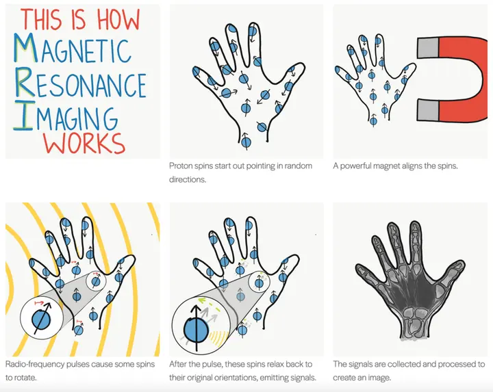
Subsequently, when a Radio Frequency (RF) pulse is applied at the appropriate resonance frequency, these protons absorb energy and their magnetic orientation tilts by 90 degrees. As soon as the RF pulse is turned off, the protons begin to return to their original equilibrium state, and during this process, they emit energy in the form of radio waves. These signals are captured by antennas or receiving coils placed nearby.
- 1. Static magnetic fieldPlacing the patient in a powerful main magnet aligns the body's hydrogen protons with the magnetic axis.
- 2. RF pulse transmissionApplying radio waves at a specific resonance frequency causes protons to absorb energy and change their orientation.
- 3. Gradient localizationTurning on gradient coils allows for 3D localization of specific field areas.
- 4. Relaxation and signal acquisitionWhen the RF pulse is turned off, protons return to their original state and the emitted RF signal is captured by an antenna.
- 5. Computer imagingThe computer analyzes these signals and phase changes to create a sharp image of body parts.
The process by which protons return to their equilibrium state after the RF pulse is turned off is called relaxation. This relaxation occurs through two independent processes: T1 (spin-lattice) and T2 (spin-spin). T1 is the time to regain stability in the longitudinal direction, while T2 is the time for the signal to decay to zero in the transverse plane. These two rates are defined as R1 = 1/T1 and R2 = 1/T2, and their behavior is determined by the Bloch equations.
T1 and T2 times vary according to the chemical environment of hydrogen in different tissues. For example, the cerebral cortex or fatty regions appear clearly in T1-weighted images, while swelling, inflammation, or fluid are more accurately visible in T2-weighted images. By adjusting the scanner's timing parameters (TR and TE), doctors obtain the necessary image contrast.
| Feature | T1-weighted image | T2-weighted image |
|---|---|---|
| Main time parameter | Repetition Time (TR) | Echo Time (TE) |
| Relaxation type | Spin-lattice (longitudinal) | Spin-spin (transverse) |
| Appearance of fat and fluid | Fat bright, fluid dark | Fluid/water appears bright |
| Main medical use | Brain cortex and fatty tissues | Swelling, inflammation, and white matter lesions |
The main components of an MRI scanner include the main magnet, shim coils, gradient system (gradient coils), and the RF system. The gradient coils serve to determine spatial localization in all three dimensions. During scanning, these coils switch on and off extremely rapidly, which generates a loud, distinctive noise from the MRI machine due to magnetostriction.
The strength of the magnet is measured in the unit Tesla (T). Most clinical scanners have a 1.5T capacity, but 3T systems are considered optimal for clearer images. In the research field, machines up to 9.4T, 10.5T, and 11.7T are used, while 14T plans are under design. Most magnets are superconducting magnets, which use liquid helium to keep them cooled to extremely low temperatures.
For patients who suffer from claustrophobia, "open" MRI designs featuring permanent magnets have been developed. Additionally, a portable MRI scanner was approved by the FDA in 2020. Recently, MRI has also become possible in the ultra-low field range of microtesla to millitesla using SQUID (Superconducting Quantum Interference Devices) technology.
Compared to CT scans, MRI provides excellent images of soft tissues such as the brain and spinal cord. In neurology, MRI is the first choice for the diagnosis of multiple sclerosis, meningitis, Alzheimer's, brain tumors, and epilepsy. It is also extremely useful for the staging of rectal and prostate cancer.

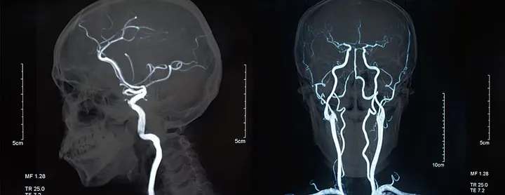
Magnetic Resonance Angiography (MRA) is used to study blood vessels in the body and brain, while functional MRI (fMRI) can determine brain activity based on blood circulation. This technology is also used for scanning inanimate objects such as mummies.
In modern times, artificial intelligence (AI) and deep learning have led to significant improvements in the speed of MRI. The fastMRI project, which began in 2018 between Meta AI Research (FAIR) and NYU Langone Health, demonstrated that with the help of AI-based image reconstruction, scanning times for knees and brains can be accelerated by a factor of 4.