Micro-CT Scanning: A Modern 'X-ray' Vision Unlocking the Secrets of Historical Artifacts Without Breaking Them
From a 443-year-old lock to space rocks, how micro-CT technology opens the doors to their internal worlds without causing any damage.
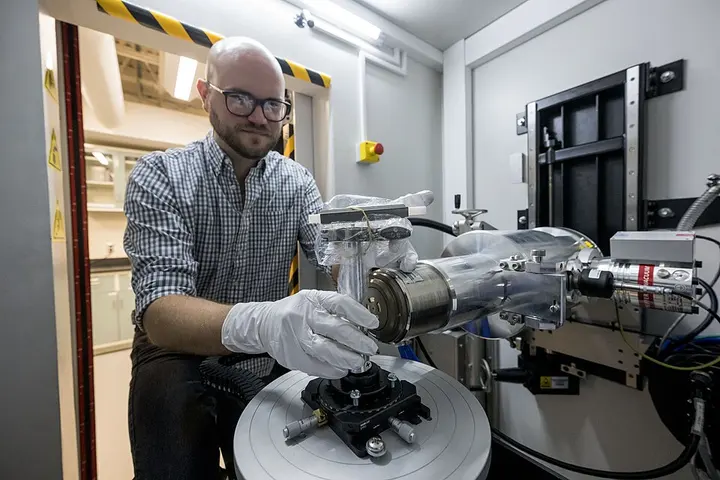
At the intersection of archaeology and science, a technology has emerged that grants the power to peek inside the closed doors of history without physically opening them. Recently, from the wreck of a 1583 ship found off the coast of Wales, researchers recovered an ancient brass combination lock. This lock had been buried in the depths of the sea for the past 443 years, and its internal parts had jammed due to rust and time. Had an attempt been made to force this lock open, it would have been destroyed forever. However, with the help of micro-CT scanning, scientists were able to view the internal structure of the lock without even touching it and also decode its four-letter secret code.
This success is not limited to just one lock; rather, it demonstrates how modern science can safely unearth the secrets of the past. Computed Tomography or CT scan, previously known as Computed Axial Tomography (CAT scan), is a method originally developed for medical diagnosis. Using this technology, detailed images of the internal parts of the body are obtained. Following its development in the 1970s, CT scanning has proven to be an extremely versatile imaging technique. British electrical engineer Godfrey Hounsfield and South African-born American physicist Allan MacLeod Cormack were awarded the 1979 Nobel Prize in Physiology or Medicine for this revolutionary invention.
- X-ray projectionA rotating tube projects X-ray beams at various angles onto the object.
- Absorption measurementX-ray beams are attenuated according to the internal density of the object, which is measured by detectors.
- Data processingA computer combines these thousands of measurements and processes them using mathematical algorithms.
- Virtual slice creationThin slices or a 3D model of the interior of the object are generated.
It is fascinating to understand how a CT scanner works. It consists of a rotating X-ray tube and a row of detectors arranged in a circular structure called a gantry. When an object or person is placed inside this machine, X-rays pass through it from various angles. Different parts of the object absorb X-ray rays in varying amounts, which is called X-ray attenuation. These measurements are processed on a computer using tomographic reconstruction algorithms. As a result, virtual 'slices' or cross-sectional images of the interior of the object are generated. This process is akin to being able to view each individual slice inside a whole loaf of bread without actually cutting it.
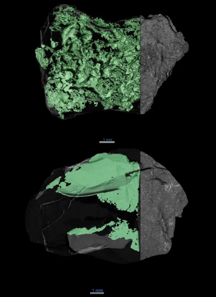
Micro-CT is a more powerful form of medical CT scans. Where a standard medical scanner provides details at the millimeter level, micro-CT can reveal the internal structure of an object at the micrometer level—smaller even than the thickness of a hair. This is why space agencies like NASA use it to examine meteorite or asteroid samples. According to a Wikimedia Commons report, X-ray computed tomography scans make it possible to clearly see the network of internal cracks within an asteroid sample, providing vital information about its formation and history.
| Type | Main Feature | Use |
|---|---|---|
| Sequential CT | Step-and-shoot method | Scanning stationary organs |
| Spiral CT | Continuous rotating tube and table | Fast and affordable scanning |
| Electron Beam (EBCT) | Extremely fast sweep speed | Heart and artery examination |
| Dual Energy CT | Two different energy levels | Density and chemical distinction of materials |
| PET-CT | Hybrid imaging | Cancer detection and metabolic activity |
There are several types of CT scanners based on technology. The most common type is 'sequential CT', in which the scanning table moves forward step-by-step and a slice is taken at each stop. On the other hand, in 'spiral CT' or helical CT, the X-ray tube rotates continuously while the table also moves continuously, facilitating faster scanning. For dynamic structures like the heart and arteries, 'electron beam computed tomography' (EBCT) is used, which prevents blurring in the images due to its extreme speed. Additionally, 'dual energy CT' uses two different energy levels to provide more precise information about a material's density and chemical composition.
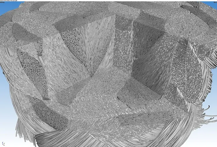
The use of micro-CT is also increasing in industries. For example, it is used to examine the internal structure of polymer ropes. 3D micro-CT scan renders from Wikimedia Commons show how the internal fibers of a rope are woven together. This type of scanning helps understand the strength of the rope and any flaws within it, which is critical for mountaineering or industrial safety. With the help of this technology, we can view the internal weave and cross-section of an object in detail without damaging its external appearance.
The use of CT scans in the medical field has increased dramatically. In the United States, approximately 72 million scans were performed in 2007, which increased to over 80 million by 2015. Head CT scans are essential for detecting strokes, tumors, calcium deposits, or bone injuries in the brain. In the case of lungs, abnormalities that standard X-rays cannot show can be detected through high-resolution CT. However, frequent scanning also carries radiation risks. Sometimes 'incidentalomas', which are small, symptomless nodules, are found in the lungs; while mostly harmless, they cause unnecessary anxiety among patients and increase the tendency for repeated scans.
કરોડ સ્કેન
CT scans are considered the gold standard for complex bone and joint injuries because they can reveal details as fine as 0.2 millimeters. For cardiac examinations, 'Coronary CT Angiography' (CCTA) is used, in which a contrast agent is injected into the veins to check for blockages in the arteries. In addition, non-contrast CT scans are the most accurate method for diagnosing kidney stones, helping to determine treatment by measuring the size, dimensions, and density of the stones.
For a country like India, which has a history thousands of years old and countless archaeological remains, micro-CT technology can prove to be a blessing. Our ancient manuscripts that risk breaking the moment they are opened, or metal objects excavated from the earth that have fused together due to corrosion, can be digitally unwrapped using this technology. For the general public, this technology means that in the future we will be able to experience history not just from the outside, but along with the secrets hidden within it. However, the cost of high-quality micro-CT scanners and the availability of experts required to operate them remain a challenge, but as the technology becomes more affordable, its scope will continue to expand.